Structure and Function of a cell
Introduction —What is Physiology?
The term physiology is originally derived from a Greek root with Latin equivalent physiologia which denotes natural knowledge. The knowledge of physiology is important to appreciate the role of mechanism that controls bodily functions.
Physiology is, therefore, the discipline that deals with the bodily functions and their control. It is however, only, concerned with the normal bodily functions and their control.
Homeostatic Regulation
The cells, tissues, organs and organ systems of the body are interconnected and live together in a shared (internal) environment. Blood forms the internal environment of the cell, ie. millieu interieur in terms of volume, composition, concentration, pH and temperature. This is regulated to normal physiological limits with respect to minor changes in the body.
A variety of physiological mechanisms which act to stabilise the internal environment, are called homeostasis. The adjustments in physiological systems, those are responsible for the preservation of homeostasis, are referred to as homeostatic regulation.
Homeostatic regulation usually involves a receptor, sensitive to a particular stimulus, and an effector whose activity affects the same stimulus.
A. Negative Feedback Mechanisms
Most homeostatic mechanisms involve negative feedback, i.e. a corrective mechanism involving an action that directly opposes a variation from normal limits. For example:
Rise in blood pressure ————————————————> Decrease in BP to normal
( a stimulus ) —————— → via -ve feedback mechanism ———– ( response )
Important note
Here the initial stimulus produces a response that depresses the stimulus, i.e. stimulus and response are opposite to each other.
In general, the nervous system performs corrective management by directing rapid, short-term and very specific responses. On the other hand, the endocrine system releases chemical messengers (hormones) that affect tissues and organs throughout the body. The response may be slow to begin with but often persists for days or weeks. However, both systems are usually controlled by negative feedback mechanisms.
B. Positive Feedback Mechanisms
In few instances homeostatic regulation involves positive feedback mechanisms, ie. an initial disturbance in a system sets off a chain of events that increase the disturbance even further.
Important note
Here, the initial stimulus produces response that reinforces (exaggerates) the original stimulus.
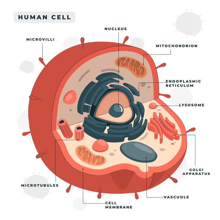
Structure and Function of a Cell
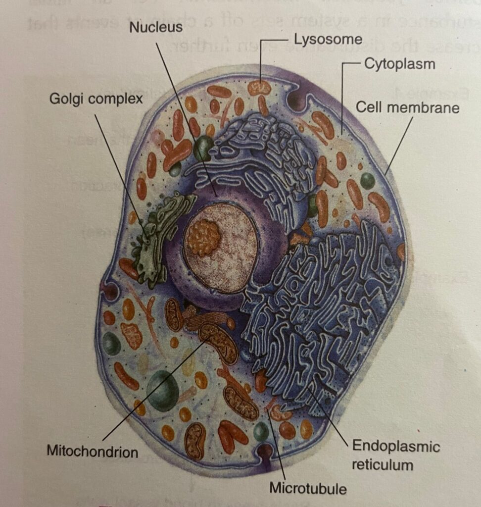
The fundamental unit of life is a cell. Most cells in a human being have diameters of 10-20 um (range 2-120 um).
The three principal constituents of a cell are:
(A) Cell membrane
(B) Nucleus and its chromosomes
(C) Cytoplasm and its organelles
Note
The clear fluid portion of the cytoplasm in which the particles are dispersed is called cytosol.
A. Cell Membrane or Plasma Membrane or Unit Membrane
Thickness
7-10 nanometer (nm) (1 nm = 10-9 m)
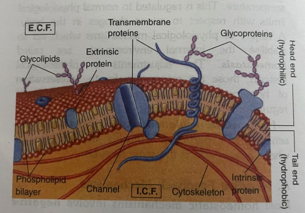
Structure (Fluid Mosaic Model)
- All membranes consist of a double layer of lipid molecules in which proteins are embedded. The lipids normally constitute 20-40% of the dry weight of the membrane.
- Proteins make up to 60-70% of the dry weight of the membrane and are of two types:
- Lipoproteins (proteins containing lipids): Function as enzymes and ion channels
- Glycoproteins (proteins containing carbohydrates constituting 1-5% of the dry weight): Function as receptors for hormones and neurotransmitters.
Some proteins are located in the inner surface of the membrane (intrinsic proteins), some are located in the outer surface of the membranes (extrinsic and peripheral proteins); while some extend through the membrane (transmembrane proteins).
(i) Intrinsic proteins serve mainly as ‘enzymes’
(ii) Extrinsic proteins contribute to the cytoskeleton (framework of the cell).
(iii) Transmembrane proteins serve as:
(a). Channels through which ions or small water soluble substances can diffuse.
(b). Carriers which passively or actively transport materials across the lipid layer.
(c). Pumps which actively transport ions across the lipid layer.
(d). Receptors which when activated initiate intracellular reactions.
- The clear area formed by bimolecular thickness of lipid molecules (phospholipids, cholesterol and glycolipids) is arranged as follows:
(i) Head end: The head end contains phosphate portion, is positively charged and quite soluble in water (i.e. polar or hydrophilic). Polar groups of lipid molecules have affinity for water (water loving) and face the aqueous phase. i.e. exterior of the cell on one side
(ECF) and cytoplasm on the other (ICF).
(ii) Tail end: The tail end is quite insoluble in water (no affinity for water/water fearing) (i.e. non-polar or hydrophobic) and contains two fatty acid chains. The hydrophobic ends facing each other meet in the water-poor interior of the membrane.
Important Note
The bimolecular lipid layer in the membrane has the characteristics of a fluid due to the presence of cholesterol. This fluidity makes the membrane quite flexible, and thus allows cells to undergo considerable changes in its shape without disruption of their structural integrity
Functions
- Protective-The cell membrane forms the outermost boundary of the cell organelles.
- Digestive—It takes in food and excretes waste products.
- Property of selective permeability
(i) Non-polar molecules (gases like 02, CO2 and N2, lipids; steroid hormones; alcohol) can dissolve in the non-polar regions of the membrane and thus move rapidly across the membrane. Polar molecules (water soluble substances: ions, glucose, urea, etc.) have much lower solubility and, therefore, penetrate the membrane much more slowly.
(ii) This property of selective permeability of the cell membrane enables to maintain the difference of composition between ECF and - It links adjacent cells together by junctional complexes to form tissues.
B. Nucleus and Its Chromosomes
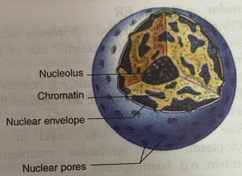
Structure
- The nucleus is a spherical structure 10 um diameter) surrounded by a relatively permeable membrane called nuclear membrane (or envelope)
- It is made up of chromosomes (each chromosome is made up of supporting protein plus giant molecule of Deoxyribonucleic Acid-DNA) on which genes are present. Gene is a portion of DNA molecule which carries a complete blue print for all the heritable species and individual characteristics of an animal. During cell division, the pairs of chromosomes become visible, but between cell divisions the irregular clumps of dark material called chromatin are the only evidence of their presence.
- It contains a nucleolus which is the densest of all the nuclear material, i.e. a patch work of granules rich in Ribonucleic Acid (RNA). It synthesizes the RNA for the ribosomes.
Functions
- DNA in nucleus serves as a ‘template’ (block) for synthesis of RNA which then moves to the cytoplasm where it regulates the synthesis of proteins by the cells.
- Genes are units of hereditary characteristics.
- It is concerned with cellular reproduction and multiplication.
C. Cytoplasm and Its Organelles
1. Endoplasmic Reticulum

The endoplasmic reticulum (ER) is a complex series of tubules whose walls are made up of unit membrane.
Through this network of tubules, substances may be delivered from the outer membrane of cell proper to the membrane of the nucleus or to cell organelles .
Types:
(i) Agranular or Smooth ER: Contains no granules.
(a) It is the site of steroid (adrenocortical hormone) synthesis in steroid secreting cells and the site of detoxification processes in other cells.
(b) As the sarcoplasmic reticulum, it plays an important role in skeletal and cardiac muscles.
(ii) Granular or Rough ER
(a) Contains granules called ribosomes which are attached to the cytoplasmic side of the membrane.
(b) It is the site of protein synthesis, e.g. hormones that are secreted by the cell; and proteins that are found in enzymes.
(c) Free ribosomes are also found in the cytoplasm. They synthesize cytoplasmic protein, e.g. haemoglobin.
2. Golgi Complex (or Golgi Bodies)

The golgi complex is a collection of membranous tubules and vesicles always found in the vicinity (neighbourhood) of the nucleus. It is prominent in actively secreting gland cells.
Functions
- Wrapping and packaging department of the cell.
- Produces secretion granules, i.e. membrane enclosed complexes which store hormones and enzymes in protein secreting cells. It also packages proteins.
- Site of formation of lysosomes (see below).
3. Mitochondrion

Structure
- (i) Length 5-12 um; diameter 0.5-1 um; filamentous or globular in shape
- (ii) Made up of outer membrane and inner membrane. Inner membrane folded to form cristae (shelves) which project into the interior of the mitochondrion.
- (iii) Outer membrane: Studded with enzymes concerned with biological oxidation (oxidation being catalyzed by enzymes).
- (iv) Interior (matrix) of mitochondrion enzymes concerned with citric acid cycle’ contains ‘respiratory chain oxidation’ and
- (v) Inner membrane is made up of repeating units each of which contains adenosine triphosphatase (ATPase) and other enzymes concerned with synthesis and metabolism of ATP.
Functions
(i) Mitochondria are the power generating units of the cells and are plentiful and best developed in parts of cells where energy requiring processes take place, e.g. rapidly contracting skeletal muscles.
(ii) Also contain DNA and can synthesize
4.Lysosomes
Structure
- Lysosomes are large irregular structures surrounded by unit membrane and are found in the cytoplasm.
They are 250-750 um in diameter. - It is filled with large number of small granules, 5-8 nm in diameter which contain variety of enzymes called lysozymes
Table : Lysosomal enzymes ( lysozymes) and the Substrates on which they act
Enzymes
- Ribonuclease
- Deoxyribonuclease
- Phosphatase
- Glycosidase
- Arylsulphatase
- Collagenase
- Cathepsins
Substrate
- RNA
- DNA
- Phosphate Esters
- Complex carbohydrate & polysaccharide
- Sulphate esters
- Proteins
- Proteins
Functions
- (i) Lysosomes act as a form of digestive (lytic) system for the cell because enzymes present in it can digest essentially all macromolecules.
- (ii) Engulf worn out components of the cells in which they are located.
- (iii) Engulf exogenous substances, e.g. bacteria, etc. and degrade them.
- (iv) When a cell dies, lysosomal enzymes cause autolysis of the remnant that is why lysosomes are called suicidal bags
5. Peroxisomes
- (i) The structure of peroxisomes is similar to that of lysosomes but with a different chemical composition. Contain oxidases rather than hydrolases)
- (ii) They destroy certain products formed from oxygen, especially hydrogen peroxide, that can be toxic to the cells. Hence, the name peroxisomes.
6.Centrioles or Centrosomes
Structure
- (i) The centrioles are two short cylinders called “centrioles’ visible only during cell division.
- (ii) They are located near the nucleus and are so arranged that they are at right angles to each other.
Function
The centrioles are concerned with the movement of the chromosomes during cell division.
7. Microtubules and Microfilaments

Microtubules are long hollow structures approximately 25 nm in diameter. They make up the structures or tracts on which chromosomes, mitochondria and secretion granules move from one part of the cell to another
Microfilaments are long solid fibres 4-6 nm in diameter. They comprise the contractile protein actin and are responsible for cell motion.
Function
The microtubules and microfilaments are involved in the
- (i) movements of the chromosomes.
- (ii) cell movement.
- (iii) processes that move secretion granules in the cell.
- (iv) movement of proteins within the cell membrane.
8. Secretion Granules
Important Note
All cells have a system of fibres called cytoskeleton that maintains the structure of the cell. It allows a cell to change shape and also permits its movement. The cytoskeleton comprises microtubules and microfilaments along with proteins that bind them together.
Junctional Complexes-Cell Junctions
The junctional complexes are associated into tissues by various means:
- Tight Junction: In tight junctions, membranes of two cells become opposed and outer layers of the membranes fuse strongly to form a barrier to the movement of ions and other solutes from
- one side of the epithelium to the other. This type of junctions are characteristically seen along the apical margins of cells in epithelium such as the intestinal mucosa, the walls of the renal tubules, and the choroid plexus.
- Desmosomes or Adherens Junction: Here two membranes are separated by 15-20 nm space.
There is dense accumulation of proteins on both the surfaces of the membrane with fibres extending from the cytoplasmic surface of each membrane into the cell. This holds adjacent cells firmly together in areas subjected to stretching, such as the skin. - Gap Junction or Nexus: There is 2 nm to 20 nm space between the opposing membranes. This gap is filled with densely packed particles through each of which there appears to be a channel that connects the two cells. The diameter of each channel is regulated by intracellular Ca?+, pH and voltage.
Other advantages of gap junction are:
(i) It permits rapid propagation of electrical potential changes from one cell to another, e.g. cardiac and smooth muscle cells.
(ii) It permits the direct transfer of ions and other small molecules upto MW 1000 (e.g. sugars, amino acids) between the cells without traversing the extracellular space.
Apoptosis: Programmed Cell Death
The word apoptosis is derived from a Greek word means loosening or falling (apo means “away’ and ptosis means ‘fall’).
- Apoptosis is a process of programmed cell death in which body cells die and get absorbed (phagocytosed) under genetic control. Here cell’s own gene plays an active role in its death.
Therefore, it is also called cell suicide. - Physiological significance: Apoptosis plays an important role during embryonal development and in adulthood. For example,
(i) it is responsible for regression of duct system during sex differentiation in the foetus. (ii) it is responsible for degeneration and regeneration of neurons within the central nervous system (CNS) and for the formation of synapse.
(iii) it is responsible for removal of inappropriate
clones of immune cells.
(iv) it is responsible for cyclical shedding of endometrium at the time of menstruation.
(v) it is responsible for cell shed from the tip of the villi in the small intestine. - Applied: Abnormal apoptosis occurs in autoimmune diseases, degenerative diseases and cancers.
To study developmental anomalies of oral cavity soft tissue structure do follow the link.
About Temporomandibular joint follow the link